Type 2 Inflammation may play a role in Prurigo Nodularis Pathophysiology1
Exact pathophysiology of PN is currently not understood. Based on the evolving literature, type 2 inflammation may play a role in PN.
.png0/jcr:content/Banner%20(1).png)
PN lesions contain a number of immune cells, including type 2 cells1-8
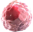
Th2 cells, Th17 cells, and Th22 cells
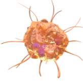
Macrophages
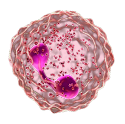
Eosinophils
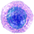
Mast cells
Basophils
Increased activity of type 2 cytokines IL-4, IL-13, and IL-31, as well as other inflammatory mediators (IL-17, IL-22, pruritogens, and neuropeptides), is present in PN skin4,5,9,10
IL-4
IL-13
IL-31
Signaling of type 2 cytokines, including IL-4 and IL-13, may contribute to signs and symptoms of PN
IL-4, IL-13, and IL-31 potential effects on itch
- Sensitization of nerves to pruritogens11,a
- Upregulation of the receptor for IL-3112,13
IL-4, IL-13, and IL-31 potential effects on skin
- Increased fibroblast proliferation and collagen production,periostin expression and stimulating profibrotic cytokine TGF-β production may influence skin remodeling and nodule formation11,12
- Skin barrier dysfunction: inhibition of keratinocyte differentiation and altered lipid synthesis13
- Epidermal differentiation, inflammatory responses, and extracellular remodeling14
The skin, immune system, and nervous system have a dysregulated relationship in PN15-19
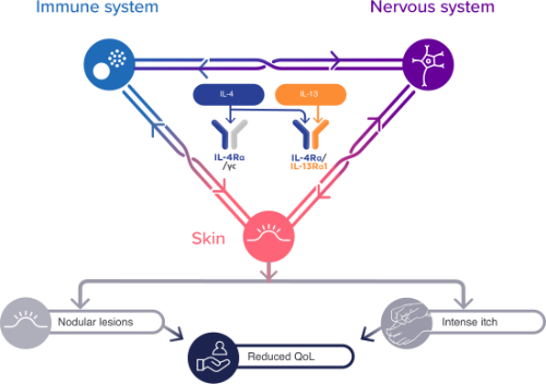
Increased activity of type 2 immune cells and cytokines, along with certain other immune cells and inflammatory mediators, may contribute to signs and symptoms of PN through interactions with neurons and other dermal and epidermal resident cells1-10,20-23
- Weigelt N, Metze D, Ständer S. Prurigo nodularis: systematic analysis of 58 histological criteria in 136 patients. J Cutan Pathol. 2010;37(5):578-586.
- Williams KA, Huang AH, Belzberg M, Kwatra SG. Prurigo nodularis: pathogenesis and management. J Am Acad Dermatol. 2020;83(6):1567-1575.
- Williams KA, Roh YS, Brown I, et al. Pathophysiology, diagnosis, and pharmacological treatment of prurigo nodularis. Expert Rev Clin Pharmacol. 2021;14(1):67-77.
- Fukushi S, Yamasaki K, Aiba S. Nuclear localization of activated STAT6 and STAT3 in epidermis of prurigo nodularis. Br J Dermatol. 2011;165(5):990-996.
- Park K, Mori T, Nakamura M, Tokura Y. Increased expression of mRNAs for IL-4, IL-17, IL-22 and IL-31 in skin lesions of subacute and chronic forms of prurigo. Eur J Dermatol. 2011;21(1):135-136.
- Ito Y, Satoh T, Takayama K, Miyagishi C, Walls AF, Yokozeki H. Basophil recruitment and activation in inflammatory skin diseases. Allergy. 2011;66(8):1107-1113.
- Nakashima C, Ishida Y, Kitoh A, Otsuka A, Kabashima K. Interaction of peripheral nerves and mast cells, eosinophils, and basophils in the development of pruritus. Exp Dermatol. 2019;28(12):1405-1411.
- Hashimoto T, Rosen JD, Sanders KM, Yosipovitch G. Possible roles of basophils in chronic itch. Exp Dermatol. 2019;28(12):1373-1379.
- Sonkoly E, Muller A, Lauerma AI, et al. IL-31: a new link between T cells and pruritus in atopic skin inflammation. J Allergy Clin Immunol. 2006;117(2):411-417.
- Fostini AC, Girolomoni G, Tessari G. Prurigo nodularis: an update on etiopathogenesis and therapy. J Dermatolog Treat. 2013;24(6):458-462.
- Oetjen LK, Mack MR, Feng J, et al. Sensory neurons co-opt classical immune signaling pathways to mediate chronic itch. Cell. 2017;171(1):217-228.e13.
- Miake S, Tsuji G, Takemura M, et al. IL-4 augments IL-31/IL-31 receptor alpha interaction leading to enhanced Ccl 17 and Ccl 22 production in dendritic cells: implications for atopic dermatitis. Int J Mol Sci. 2019;20(16):4053.
- Hashimoto T, Nattkemper LA, Kim HS, et al. Dermal periostin: a new player in itch of prurigo nodularis. Acta Derm Venereol. 2021;101(1):adv00375.
- Tsoi LC, Hacini-Rachinel F, Fogel P, et al. Transcriptomic characterization of prurigo nodularis and the therapeutic response to nemolizumab. J Allergy Clin Immunol. Published online November 29, 2021. doi:10.1016/j.jaci.2021.10.004.
- Mack MR, Kim BS. The itch-scratch cycle: a neuroimmune perspective. Trends Immunol. 2018;39(12):980-991.
- Zeidler C, Yosipovitch G, Ständer S. Prurigo nodularis and its management. Dermatol Clin. 2018;36(3):189-197.
- Kwatra SG. Breaking the itch-scratch cycle in prurigo nodularis. N Engl J Med. 2020;382(8):757-758.
- Janmohamed SR, Gwillim EC, Yousaf M, Patel KR, Silverberg JI. The impact of prurigo nodularis on quality of life: a systematic review and meta-analysis. Arch Dermatol Res. 2021;313(8):669-677.
- Pereira MP, Hoffmann V, Weisshaar E, et al. Chronic nodular prurigo: clinical profile and burden. A European cross-sectional study. J Eur Acad Dermatol Venereol. 2020;34(10):2373-2383.
- Campion M, Smith L, Gatault S, Métais C, Buddenkotte J, Seinhoff M. Interleukin-4 and interleukin-13 evoke scratching behaviour in mice. Exp Dermatol. 2019;28(12):1501-1504.
- Edukulla R, Singh B, Jegga AG, Sontake V, Dillon S, Madala S. Th2 cytokines augment IL-31/IL-31RA interactions via STAT6-dependent IL-31RA expression. J Biol Chem. 2015;290(21):13510-13520.
- Nguyen JK, Austin E, Huang A, Mamalis A, Jagdeo J. The IL-4/IL-13 axis in skin fibrosis and scarring: mechanistic concepts and therapeutic targets. Arch Dermatol Res. 2020;312(2):81-92.
- Serezani APM, Bozdogan G, Sehra S, et al. IL-4 impairs wound healing potential in the skin by repressing fibronectin expression. J Allergy Clin Immunol. 2017;139(1):142-151.e5.
Mechanism of Prurigo Nodularis Disease
In this comprehensive exploration, we delve into the intricate pathophysiology of Prurigo Nodularis (PN), highlighting the pivotal roles of neuronal sensitization, inflammation, and pruritus. This challenging dermatological condition is characterized by dermatological condition histopathologic alterations, including enhanced hypervascularization, epidermal hyperkeratosis, and dermal fibrosis. Explore the connection between PN and the proinflammatory Th2 cytokine IL-13, shedding light on its binding to IL-4R across various skin components.
