{ event: "article_read", name: `Differential Diagnosis Of Late Onset Pompe Disease (LOPD) and Myotonic Dystrophy Type 1 Through Abdominal Ultrasonography`, author: ``, tags: `Rare diseases`, publication_date: ``, interaction_type: "content" } 
Differential Diagnosis Of Late Onset Pompe Disease (LOPD) and Myotonic Dystrophy Type 1 Through Abdominal Ultrasonography
.png/jcr:content/science%20hero%20(1).png)
Study objective and method
- Compare ultrasonography images
- In patients with LOPD (n=3), DM1 (n=10), and age- and gender-matched healthy subjects (n=34)
- Muscle thickness and echogenicity were assessed
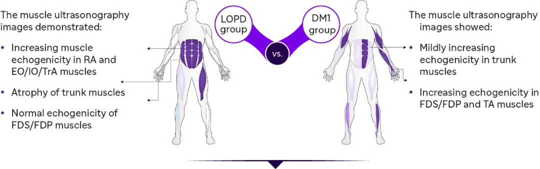
Results
Qualitative echogenicity in muscular ultrasonography
The variable severity and distribution of qualitative echogenicity (Z scores) in skeletal muscles of patients with LOPD and DM1
Qualitative echogenicity in skeletal muscles:
- Abnormal in the abdominal muscles
- Mildly abnormal in rectus femoris and anterior tibialis muscles
| Z Score | ||
| Normal | <2 | |
| Mild | 2-4 | |
| Moderate | >4 | |
| Severe | >6 |
Qualitative echogenicity in skeletal muscles:
- Abnormal in abdominal muscles and more obvious in biceps, TA, and FDS/FDP
Quantitative echogenicity and abdominal muscle thickness in muscular ultrasonography

Quantitative muscle echogenicity

Trunk muscle thickness
.jpg/jcr:content/Asset%208@3x%20(1).jpg)
Total trunk grading sum score
Conclusion
These findings suggest that muscle ultrasound is an efficient screening tool for:
- Assessing myopathic changes and disease-specific patterns
- Differential diagnosis of neuromuscular diseases
- Trunk muscles can be used for the differential diagnosis of LOPD and DM1.
- Muscle echography results correlated with clinical and motor functions.
- TIS can be used to investigate the trunk function
DM1: Myotonic dystrophy type 1; EO: External oblique; FDP: Flexor digitorum profundus; FDS: Flexor digitorum superficialis; IO: Internal oblique; LOPD: Late-onset Pompe disease; RA: Rectus abdominis; TA: Tibialis anterior; TIS: Trunk impairment scale; TrA: Transversus abdominis.
- Hsieh PC, Chang CW, Ro LS, et al. Ultrasonography of abdominal muscles: Differential diagnosis of late-onset Pompe disease and myotonic dystrophy type 1. Front Neurol. 2022;13:944464.
.webp/jcr:content/gaucher-thumb%20(2).webp)


MAT-KW-2300245 V1 Jul 2023