{
event: "article_read",
name: `Differential Diagnosis Of Late Onset Pompe Disease (LOPD) and Myotonic Dystrophy Type 1 Through Abdominal Ultrasonography`,
author: ``,
tags: `Rare Diseases | Rare Diseases`,
publication_date: ``,
interaction_type: "content"
}

Read article
Read article
Differential Diagnosis Of Late Onset Pompe Disease (LOPD) and Myotonic Dystrophy Type 1 Through Abdominal Ultrasonography
.jpg/jcr:content/jcr_content%20(38).jpg)
Study objective and method
- Compare ultrasonography images
- In patients with LOPD (n=3), DM1 (n=10), and age- and gender-matched healthy subjects (n=34)
- Muscle thickness and echogenicity were assessed
Results
Qualitative echogenicity in muscular ultrasonography
%20(1).2024-03-18-09-01-03.jpg)
The variable severity and distribution of qualitative echogenicity (Z scores) in skeletal muscles of patients with LOPD and DM1
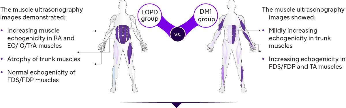
Qualitative echogenicity in skeletal muscles:
- Abnormal in the abdominal muscles
- Mildly abnormal in rectus femoris and anterior tibialis muscles
| Z Score | ||
| Normal | <2 | |
| Mild | 2-4 | |
| Moderate | >4 | |
| Severe | >6 |
Qualitative echogenicity in skeletal muscles:
- Abnormal in abdominal muscles and more obvious in biceps, TA, and FDS/FDP
Quantitative echogenicity and abdominal muscle thickness in muscular ultrasonography
.jpg/jcr:content/Asset%2010@3x%20(1).jpg)
Quantitative muscle echogenicity
.jpg/jcr:content/Asset%209@3x%20(3).jpg)
Trunk muscle thickness
-(3).jpg/jcr:content/Asset%208@3x%20(1)%20(3).jpg)
Total trunk grading sum score
Conclusion
These findings suggest that muscle ultrasound is an efficient screening tool for:
- Assessing myopathic changes and disease-specific patterns
- Differential diagnosis of neuromuscular diseases
- Trunk muscles can be used for the differential diagnosis of LOPD and DM1.
- Muscle echography results correlated with clinical and motor functions.
- TIS can be used to investigate the trunk function
DM1: Myotonic dystrophy type 1; EO: External oblique; FDP: Flexor digitorum profundus; FDS: Flexor digitorum superficialis; IO: Internal oblique; LOPD: Late-onset Pompe disease; RA: Rectus abdominis; TA: Tibialis anterior; TIS: Trunk impairment scale; TrA: Transversus abdominis.
- Hsieh PC, Chang CW, Ro LS, et al. Ultrasonography of abdominal muscles: Differential diagnosis of late-onset Pompe disease and myotonic dystrophy type 1. Front Neurol. 2022;13:944464.
Related articles

.jpg/jcr:content/jcr_content%20(37).jpg)
Expert Group Consensus From The Arabian Peninsula On The Diagnosis Of Late Onset Pompe Disease for orthopedists
.jpg/jcr:content/jcr_content%20(35).jpg)
Expert Group Consensus From The Arabian Peninsula On The Diagnosis Of Late Onset Pompe Disease (LOPD)
MAT-KW-2300245 V1 Jul 2023